

Multimodal molecular imaging in medical monitoring, diagnosis, and treatment
Dr Md Anawar Hossain
Molecular imaging
Molecular imaging is a powerful interdisciplinary imaging technology that performs the in vivo imaging and describes the molecular biology of human body organs (Wu and Shu, 2018). This technique can depict and elucidate living biological processes at the tissue, cellular and molecular level in a non-invasive manner. It can also monitor and diagnose disease changes and monitor human health. Molecular imaging can detect the abnormal changes in cells and molecules causing the diseases and help to identify the early tumor diagnosis and indication of diseases.
They can present images of the internal body organs, tissue, and cells, which describe, predict and indicate the current state and future trend of health conditions. They can significantly help the healthcare professionals to diagnose the current state and prognosis of disease, and accordingly can take the steps of treatment processes.
Traditional and molecular imaging
Traditional imaging techniques can’t detect the living biological process at the cellular and molecular level. By contrast, molecular imaging can identify the changes in the cellular and molecular levels that can cause diseases. It can do the early diagnosis and a more accurate diagnosis of the disease (Liu et al., 2022).
Types of molecular imaging techniques
Different molecular imaging technologies have nowadays been used to obtain in vivo imaging and identify the structural, functional, cellular, and molecular changes in body organs, tissues and tumor tissues. These techniques include optical imaging (either by bioluminescence or fluorescence), computed tomography (CT), magnetic resonance imaging (MRI), positron emission tomography (PET), single-photon emission computed tomography (SPECT), and ultrasound (US). These techniques are importantly used in clinical research, and for patients.
Multimodal molecular imaging
Different types of molecular imaging techniques are currently under use. These techniques have their own advantages, disadvantages, and limitations in terms of the spatial/depth resolution and sensitivity, making the achievement of precise and reliable information at the disease site. Therefore, we need to develop a more advanced and high-performance imaging technique to overcome these disadvantages and limitations. Researchers have combined two or more imaging techniques to create a new imaging system, such as multimodal molecular imaging for obtaining some further information in diagnosis, treatment, and monitoring. Wu and Shu (2018) discussed the benefits of the classic molecular imaging technology and proposed some of the latest multimodal molecular imaging modes.
Benefit of multimodal molecular imaging
- Multimodal molecular imaging is presumed to produce a better result and more accurate information regarding monitoring, diagnosis, and treatment of difficult diseases.
- This technique can help the healthcare professionals to perform screening, surveillance, staging, prognosis, planning and therapy guidance, monitoring therapy efficacy, and assessing recurrence.
- It may also aid in presymptomatic detection, targeted therapy, and personalized medicine.
- It has shown promising results in the treatment of cardiovascular diseases, neuropsychiatric diseases, and other clinical diseases.
- It can significantly enhance the positioning of the tumor border and effectively guide the surgical resection of the tumor.
A few specific examples are as follows:
Cholangiocarcinoma (CCA):
Molecular imaging methods such as nuclear medicine imaging (PET and SPECT), MRI, optical imaging, and multimodal imaging are.used in clinical and preclinical research in cholangiocarcinoma (CCA), which is an aggressive and lethal malignant tumor invasion into the bile duct and its surrounding tissues (Liu et al., 2022).
Cardiovascular diseases:
Yoo (2018) combined coherence tomography and fluorescence lifetime imaging to form multimodal intravascular optical imaging which can provide new opportunities to investigate vascular pathobiology and diagnose cardiovascular disease, by simultaneously visualizing plaque morphology and biochemical composition.
Neuropsychiatric diseases:
Voss et al. (2011) combined functional MRI and PET to form multimodal functional-imaging technology and studied a patient with marked neurological recovery after cranioplasty.
Other clinical diseases:
Tang et al. (2016) developed a novel multimodal video endoscope and evaluated its usefulness for the early detection of gastric neoplastic lesions.
Molecular Imaging Models
Most important and widely used molecular imaging models are discussed below:
Computed tomography (CT) molecular Imaging
CT is a widely available technique which can provide images of human body organs reflecting human anatomy.
Advantages
- It has high spatial resolution, strong penetration depth, fast acquisition time, low cost, clinical utility, and relative simplicity.
- It can detect and assess several diseases, such as tumors, brain injury, and pulmonary embolism.
- It is a useful morphological tool.
Disadvantages
- It has high radiation dose, which often limits the scan time.
- Low-quality soft tissue contrast, compared with MRI.
- Low sensitivity of current CT contrast agents.
- It needs high quality imaging agents, which are under continuing research.
Magnetic resonance imaging (MRI) molecular imaging
MRI is the most useful imaging technique in radiology, especially for the detection and characterization of soft tissue pathology.
Advantages
- MRI can provide three-dimensional clear images.
- It has high spatial resolution and high contrast.
- The acquisition time and the quality of images are constantly improving.
- It is one of the most important molecular imaging research methods.
- It can be used for non-invasive monitoring and early diagnosis of diseases at the cellular and molecular level.
- The study of MRI is increasing for cell tracking, angiogenesis, apoptosis, and in vivo tissue gene imaging.
Disadvantages
The techniques still have some difficulties requiring an urgent solution.
Radionuclide Molecular Imaging
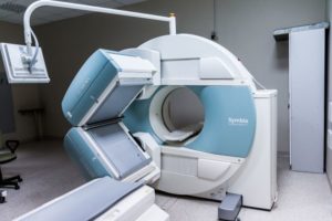
Radionuclide imaging is a radioactive marker in drug. SPECT and PET are advanced radionuclide molecular imaging techniques. Both techniques enable whole body imaging of molecular targets/processes with high sensitivity.
SPECT is mainly used for whole body bone imaging, myocardial blood flow imaging, cerebral blood flow imaging, and thyroid imaging.
PET is widely used for the nervous system, the cardiovascular system, and the oncology.
PET and SPECT can identify and indicate the treatment of various diseases and tumors.
Ultrasound Molecular Imaging
Ultrasound imaging provides a morphological imaging similar to MRI and CT. Medical ultrasound imaging uses the properties and behavior of high-frequency sound waves as they travel through biological tissue.
Advantages
- It can be used both for diagnostic imaging and as a therapeutic tool.
- Ultrasound imaging is better than traditional imaging techniques such as radionuclide imaging and optical imaging in terms of economy, convenience, and real-time imaging.
- It can present the early diagnosis and specific treatment of malignant tumors.
Optical molecular imaging
Optical imaging uses optical detection means combined with optical detection molecules to imaging cells or tissues or even organisms. There are several biological optical imaging methods, such as fluorescence imaging, bioluminescence imaging, photoacoustic imaging, and optical tomography. Optical imaging has still now some defects in respect of the living organism imaging. It is currently used in the basic and applied medical research using small animal and needs further research for improvement.
Fluorescence molecular tomography (FMT)
This technique can visualize and quantitate a variety of cellular and molecular events. FMT is different from planar fluorescence imaging. FMT can produce quantitative information and allows imaging at greater depths, up to several centimeters.
Bioluminescence imaging technology
This technique uses luciferase gene to label cells or DNA and exploits sensitive optical detection instrument to directly monitor cell activity and gene behavior in living subjects.
Advantages:
* Noninvasive,
* Continuous repeated detection,
* Fast real-time scanning imaging, and
* High sensitivity.
Photoacoustic imaging (PA)
Advantage
- It is a recently developed nondestructive imaging method.
- It combines the high penetration characteristics of pure optical imaging and high contrast characteristics by light into the ultrasound.
- It can provide tissue imaging with high resolution and high contrast.
References
Liu J., Ren, W.X., Shu J., 2022. Multimodal molecular imaging evaluation for early diagnosis and prognosis of cholangiocarcinoma. Insights Imaging 13, 10. Creative Commons Attribution 4.0 International License.
Tang Y., Carns J., Quang T., et al., 2016. A multimodal optical imaging platform for the early detection of gastric malignancies. Journal of Global Oncology, vol. 2, no. 3, p. 6.
Voss H.U., Heier L.A., Schiff N.D., 2011. Multimodal imaging of recovery of functional networks associated with reversal of paradoxical herniation after cranioplasty. Clinical Imaging, vol. 35, no. 4, p. 253.
Wu M., Shu J., 2018. Multimodal molecular imaging: current status and future directions. Contrast Media & Molecular Imaging Vol. 2018, Article ID 1382183, 12 pages. Creative Commons Attribution License.
Yoo H., 2018. Label-free multimodal intravascular optical imaging for cardiovascular disease, in Proceedings of Biophotonics Congress: Biomedical Optics Congress, Bangkok, Thailand, May 2018.